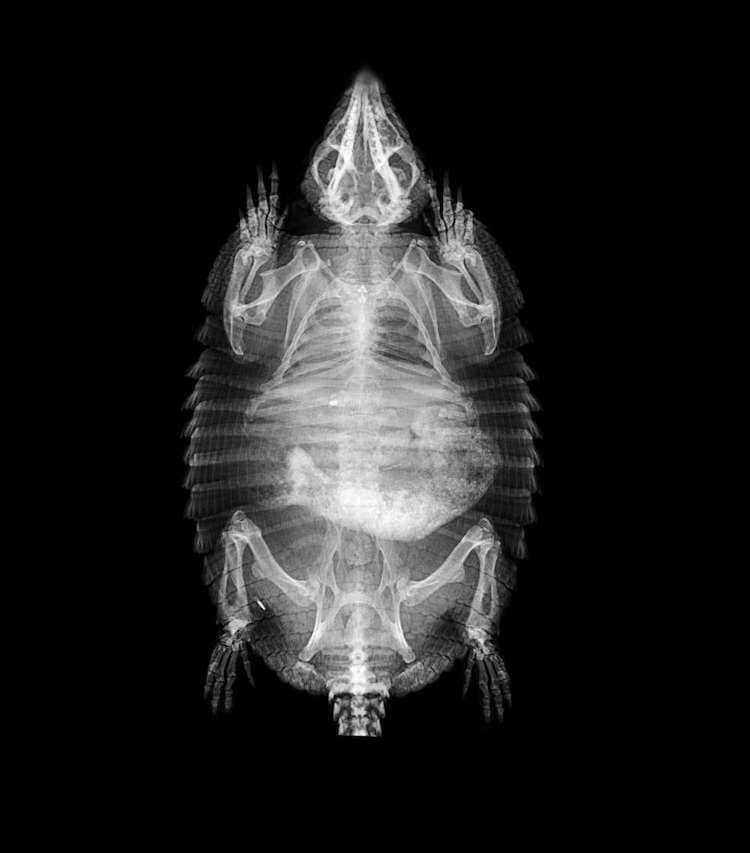
The Animal X Ray. All veterinary professionals should practice simple methods of keeping exposure as low as reasonably achievable (alara), such as. Thus, the nearer the object under examination is to the film, the sharper will be its outline. Ba, rvt | purdue university. A fundamental understanding of how radiographs are created enables the user to select the most appropriate exposure factor settings during radiograph production, in order to achieve optimal image quality. The body’s soft tissues do not absorb x‑rays well and can be difficult to see using this technology alone. Radiography is a commonly used diagnostic tool in veterinary practice. In these procedures the animal is given a dye that will block x‑rays. Because a radiograph is, in essence, a shadowgraph, the geometric rules applicable to the formation of shadows are also valid for radiographs. Specialized x‑ray techniques, called contrast procedures, are used to help provide more detailed images of body organs.
from mymodernmet.com
The body’s soft tissues do not absorb x‑rays well and can be difficult to see using this technology alone. Ba, rvt | purdue university. Thus, the nearer the object under examination is to the film, the sharper will be its outline. All veterinary professionals should practice simple methods of keeping exposure as low as reasonably achievable (alara), such as. In these procedures the animal is given a dye that will block x‑rays. Specialized x‑ray techniques, called contrast procedures, are used to help provide more detailed images of body organs. A fundamental understanding of how radiographs are created enables the user to select the most appropriate exposure factor settings during radiograph production, in order to achieve optimal image quality. Because a radiograph is, in essence, a shadowgraph, the geometric rules applicable to the formation of shadows are also valid for radiographs. Radiography is a commonly used diagnostic tool in veterinary practice.
London Zoo Releases Fascinating Animal XRays to the Public
The Animal X Ray Thus, the nearer the object under examination is to the film, the sharper will be its outline. A fundamental understanding of how radiographs are created enables the user to select the most appropriate exposure factor settings during radiograph production, in order to achieve optimal image quality. Specialized x‑ray techniques, called contrast procedures, are used to help provide more detailed images of body organs. Radiography is a commonly used diagnostic tool in veterinary practice. Ba, rvt | purdue university. Because a radiograph is, in essence, a shadowgraph, the geometric rules applicable to the formation of shadows are also valid for radiographs. All veterinary professionals should practice simple methods of keeping exposure as low as reasonably achievable (alara), such as. In these procedures the animal is given a dye that will block x‑rays. The body’s soft tissues do not absorb x‑rays well and can be difficult to see using this technology alone. Thus, the nearer the object under examination is to the film, the sharper will be its outline.